 1861
1861
 2023-02-27
Partial Spectrum Detection and Super-Gaussian Window Function for Ultrahigh-resolution Spectral-domain Optical Coherence Tomography with a Linear-k Spectrometere
Hyun-Ji Lee1,2 and Sang-Won Lee1,2 *
Current Optics and Photonics Vol. 7 No. 1 (2023 February) pp. 73-82
DOI: https://doi.org/10.3807/COPP.2023.7.1.73
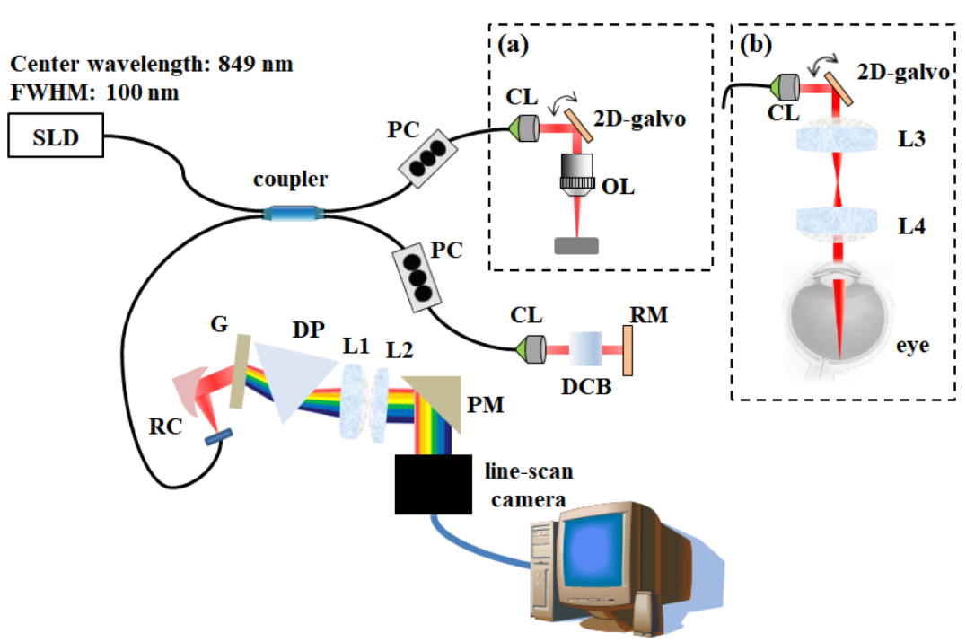
Fig. 1 Schematic of the SD-OCT based on a linear k-domain spectrometer. (a) The sample arm to measure the system performance and obtain the skin tissue’s image. (b) The sample arm to get the image of the retina. SD-OCT, spectral domain optical coherence tomography; PC, polarization controller; CL, collimation lens; OL, objective lens; DCB, dispersion compensation block; RM, reference mirror; RC, reflective collimator; G, grating; DP, dispersive prism; PM, prism mirror; L1 to L4, lenses.
글쓴이와 관리자만 접근할 수 있습니다.
글 등록 시 입력한 비밀번호를 입력해 주세요.
'한국광학회' 은 (이하 ' 한국광학회 ') 고객님의 개인정보를 중요시하며, "정보통신망 이용촉진 및 정보보호"에 관한 법률을 준수하고 있습니다. 한국광학회는 개인정보취급방침을 통하여 고객님께서 제공하시는 개인정보가 어떠한 용도와 방식으로 이용되고 있으며, 개인정보보호를 위해 어떠한 조치가 취해지고 있는지 알려드립니다.
본 방침은 : 2021 년 4 월 8 일 부터 시행됩니다.
기타 개인정보침해에 대한 신고나 상담이 필요하신 경우에는 아래 기관에 문의하시기 바랍니다.
This "Privacy Policy" explains the collection, use, and disclosure of "personal information" by Optics and Photonics Congress 2021, hosted by Optical Society of Korea (OSK hereafter), through the Website that OSK operates at osk.or.kr. In accordance to the Information Network Promotion Law revised in February 19, 2016 and legislated to prevent the misuse and abuse of personal information on the Internet, this Privacy Policy also explains our commitment to you with respect to our use and disclosure of the personal information that OSK collects from its members. That commitment is contained in a "Data Usage Policy," below. As used in this policy, "personal information" means information that would allow a party to identify you such as, for example, your name, address or location or email address. OSK collects membership information in order to provide better services to its users. By accessing osk.or.kr as your ID, you are accepting and agreeing to the practices described in this Privacy Policy.
We collect information solely for the purpose of providing services to our members and facilitating our communication with them. We do not collect or use personal information for any other purposes. The personal information provided to us by the members makes it possible for us to deliver, based on it, more useful information selectively.
Emails and Newsletters. We use the personal information you provide to us when you receive our newsletter in order to respond to your request, to reply to your email or to send you communications about news and events related to Optics and Photonics Congress 2021. When you subscribe to our newsletter, your name and email address is sent to and stored Optics and Photonics Congress 2021 (osk.or.kr).
Registered Users. When you register to obtain a user account on osk.or.kr, you may be asked to provide personal information to create your account and establish a password and profile. Providing your email address and/or real name is required. If you do provide your email address, it will allow us to send you your password if you should forget it. We will also use that personal information to establish and maintain your membership and provide you with the features we provide for Registered Users, and for the purpose of contacting you about upcoming news and events relevant to Optics and Photonics Congress 2021.
Any other personal information that we may collect which is not described specifically in this Privacy Policy will only be collected and used in accordance with the Principles. When the range, purpose, and use of the information collected by us change, we will make sure to notify our members and ask for their consent beforehand.
Only the minimum amount of information reasonably necessary to provide you with services should be collected and maintained, and only for so long as reasonably needed or required; This includes their name, occupational position, e-mail, postal code and address. We may collect personal information at several places on the Website,
Information you provide through our Website, or that we gather as a result of your use of our Website, should not be used for marketing or advertising purposes, nor should it be provided voluntarily (without your permission) to anyone else unless required. OSK will continue to maintain and manage its members' personal information as long as they continue to receive its services and retain their membership. However, when a member cancels his or her membership by request for de-registration, all personal information collected by us will be permanently deleted from our database, ensuring that the opening or use of such information for any purposes is no longer possible.
When our members visit our site and their web browsers ask for cookies OSK Website restricts its use of cookies only to the information that has no potential to invade their privacy, such as browser version, monitor information, operating system version, and information related to log-in.
OSK has implemented reasonable measures to protect against unauthorized access to and unlawful interception or processing of personal information that OSK stores and controls. However, no website can fully eliminate security risks. We will post a reasonably prominent notice to the Website if any such security breach occurs. User ID has security risks in addition to those described above. Among other things, User ID is vulnerable to DNS attacks, and using ID may increase the risk of phishing.
Our Data Usage Policy covers how we maintain and use information about you that is collected by our Website and server logs, including when you log into a website using your ID. Non Personal Browsing Information We Collect. When you use the Website, our servers may collect information automatically (through, for example, the use of your "IP address") about your activities while visiting the Website and information about the browser you are using. In addition, whenever you use your OSK ID to log into our Website, our server keep a log of the websites you visit.
No Linking. We do not intentionally link browsing information or information from our server logs to the personal information you submit to us. We use this information for internal purposes only, such as to help understand how the Websites are being used, to improve our Website and the features we provide, and for systems administration purposes.
No Selling or Sharing. Except in the unique situations identified in this Privacy Policy, OSK does not sell or otherwise voluntarily provide the non personal browsing information we collect about you or your website usage to third parties.
No Retention. OSK discards non personal browsing information from our server logs once we have used the information for the limited purposes noted above, under "No Linking."
Notice. If OSK is required to provide a third party with your non personal browsing information, then, OSK will use reasonable means to notify you promptly of that event, unless OSK is prohibited by law from doing so or is otherwise advised not to notify you on the advice of legal counsel.
Any other non-personal information that we collect which is not described specifically in this Privacy Policy will only be collected and used in accordance with the Principles.
The Website are not directed at children under the age of 14. We will never knowingly request personal information from anyone under the age of 14 without requiring parental consent. Our Master Terms of Use specifically prohibit anyone using our Website from submitting any personally identifiable information about persons under 14 years of age. Any person who provides their personal information to OSK through the Website represents that they are 14 years of age or older
We may occasionally update this Privacy Policy. When we do, we will also revise the Effective Date below. We encourage you to periodically review this Privacy Policy to stay informed about how we are protecting the personal information we collect. Your continued use of the Website constitutes your agreement to this Privacy Policy and any updates.
Members are responsible for the security of the passwords for their membership accounts. OSK never asks its members for their passwords, either through mail or any other methods. Under no circumstances should you ever disclose your password. We ask that you pay particular attention when you are logged in, in order to ensure that your personal information is not divulged to others.
If you have any concerns or suggestions about our Privacy Policy and management of your personal information, please do not hesitate to contact our representatives listed below through email or phone, and we will respond to you as expeditiously as possible.
If you have questions about this Privacy Policy, please contact us by email at osk@osk.or.kr
Your use of "osk.or.kr" will always be subject to, at a minimum, the terms and conditions set out in this document. These are referred to as the "Master Terms." In addition, your use of Website may also be subject to the terms of any legal notice applicable to the Website, in addition to the Master Terms. All such terms supplementing these Master Terms are referred to below as the "Additional Terms." The Master Terms, together with any Additional Terms, form a binding legal agreement between you and Optics and Photonics Congress 2021 in relation to your use of the Website. Collectively, this legal agreement is referred to below as the "Terms." If there is any contradiction between the Additional Terms and the Master Terms, then the Additional Terms shall take precedence in relation to the Website to which the Additional Terms apply. Your use of "osk.or.kr" will always be subject to, at a minimum, the terms and conditions set out in this document. These are referred to as the "Master Terms." In addition, your use of Website may also be subject to the terms of any legal notice applicable to the Website, in addition to the Master Terms. All such terms supplementing these Master Terms are referred to below as the "Additional Terms." The Master Terms, together with any Additional Terms, form a binding legal agreement between you and Optics and Photonics Congress 2021 in relation to your use of the Website. Collectively, this legal agreement is referred to below as the "Terms." If there is any contradiction between the Additional Terms and the Master Terms, then the Additional Terms shall take precedence in relation to the Website to which the Additional Terms apply.
Your access or use of osk.or.kr in any way signifies that you have read, understand and agree to be bound by the terms. By accessing or using osk.or.kr you also represent that you have the legal authority to accept the Terms on behalf of yourself and any party you represent in connection with your use of Website. If you do not agree to the Terms, you are not authorized to use Website.
Optics and Photonics Congress 2021 may change, remove, add to modify the Terms, and reserves the right to do so in its discretion. In that case, we will post the updated Master Terms or Additional Terms, as relevant, to the Website and indicate the date of revision. We may send a message to your email address, or we may display a notice on the Website indicating that the Terms have changed. All amended Terms take effect immediately. If you do not agree with any modification to the Terms, you may terminate this agreement by ceasing use of the Website. Your continued use of Website after revised Terms are effective indicate that you have read, understood and agreed to those Terms.
Optics and Photonics Congress 2021makes the Website available to you on the Terms. You may only use the Website in accordance with these Master Terms and any applicable Additional Terms. In particular but without limitation, you may not use the Website for any purpose that is unlawful or prohibited by these Master Terms, any applicable Additional Terms, or any other conditions or notices that are made available on any Website.
The Website Services are controlled and offered by Optics and Photonics Congress 2021 from its facilities in the Korea. Optics and Photonics Congress 2021 makes no representations that the Website are appropriate or available for use in other locations. If you are accessing or using any Website from other jurisdictions, you do so at your own risk and you are responsible for compliance with local law.
Users agree not to use the Website Services to:
Responsibility for Content. You understand that all material, data and information, (collectively, "Content") which you may have access to through your use of the Website are the sole responsibility of the person from which such Content originated. This includes assertions that persons may make, expressly or impliedly, about the provenance and ownership of Content that they supply, upload, list and/or link to. You acknowledge that Optics and Photonics Congress 2021 does not make any representations or warranties about the accuracy, integrity or quality of the Content made available at the instigation of users of the Website. You understand that by using the Website, you may be exposed to Content that is offensive, indecent or objectionable. Under no circumstances is Optics and Photonics Congress 2021 liable in any way for any Content, including, but not limited to: any infringing Content, any errors or omissions in Content, or for any loss or damage of any kind incurred as a result of the use of any Content posted, transmitted to, linked to or otherwise accessible or made available via the Website Services.
Content You Provide. You may only submit Content to the Website. This means that you can only submit Content that you yourself create, that is in the public domain or that you have been expressly granted the right to submit consistent with the Terms. For the avoidance of doubt, Content that infringes the rights of any third party must not be submitted. You represent, warrant and agree that no Content of any kind submitted, posted or otherwise shared by you on or through any of the Website Services, violates or infringes upon the rights of any third party, including copyright, trademark, privacy, publicity or other personal or proprietary rights, or contains libelous, defamatory or otherwise unlawful material. Further, you represent, warrant and agree not to submit any personally identifiable information. Optics and Photonics Congress 2021 may review your submissions and may delete or remove without notice any Content in its sole discretion that Optics and Photonics Congress 2021 determines violates the Terms or that may be offensive, illegal, or that might violate the rights, harm or threaten the safety of others. Optics and Photonics Congress 2021 does not endorse or support any Content posted by you. You alone are responsible for creating backup copies and replacing any Content you post on the Website, and you authorize Optics and Photonics Congress 2021 to make copies of your Content as we deem necessary in order to facilitate the posting of your Content on the Website.
Use of Content on the Website. You represent and warrant to Optics and Photonics Congress 2021 that you will use any and all Content on our Website in accordance with the applicable license. By using the Website, you agree that you are solely responsible for your use of any and all Content made available thereon. You agree that you must evaluate, and bear all risks associated with, the use of any Content, including any reliance on the provenance, ownership, accuracy, completeness, or reliability of such Content. In this regard, you acknowledge that you may not rely on any Content made available on the Website without your own independent evaluation of that Content. SNUH International Course does not guarantee that Content made available on the Website does not infringe the rights of any third party.
By registering for an account on the Website, you represent and warrant that you (1) are the age of majority in your jurisdiction or, (2) are over the age of 14 and have the express permission of a legal guardian to become a Registered User and use Services made available to Registered Users, and you further agree to abide by all of the terms and conditions of these Master Terms and any applicable Additional Terms. Optics and Photonics Congress 2021 reserves the right to modify or discontinue the accounts of Registered Users and related Services at any time. Optics and Photonics Congress 2021 disclaims any and all liability to Registered Users.
Security. You agree to (a) provide accurate, current and complete information about you, if and as may be prompted by the registration process on the any of the Website, (b) maintain the security of your password and identification, (c) maintain and promptly update your registration information and any other information you provide, and to keep it accurate and complete to, among other things, allow us to contact you, and (d) be fully responsible for all use of your account and for any actions that take place using your account. You may not set up an account or membership on behalf of another individual or entity.
Termination and Inactivation of User Accounts. Your participation as a Registered User terminates automatically upon your breach of any of these Master Terms or applicable Additional Terms. In addition, Optics and Photonics Congress 2021 may, at any time: (a) modify, suspend or terminate the operation of or access to your user account for any reason; (b) modify or change Website and Services and any applicable Terms and policies governing your user account and related Website Services for any reason; and (c) interrupt user accounts and related Website Services for any reason, all as Optics and Photonics Congress 2021 deems appropriate in its discretion. Your access to your account, and use of the Website may be terminated by you or by Optics and Photonics Congress 2021 at any time and for any reason whatsoever, without notice.
In addition, Optics and Photonics Congress 2021 reserves the right to delete and purge any account and all Content associated therewith following any prolonged period of inactivity, all as may be determined by Optics and Photonics Congress 2021 in its complete discretion.
To the fullest extent permitted by the applicable law, Optics and Photonics Congress 2021 offers the Website and Services as-is and makes no representations or warranties of any kind concerning the Website services. Optics and Photonics Congress 2021 does not warrant that the functions or content contained on the Website will be uninterrupted or error-free, that defects will be corrected, or that Optics and Photonics Congress 2021's servers are free of viruses or other harmful components. Optics and Photonics Congress 2021 does not warrant or make any representation regarding use or the result of use of the content in terms of accuracy, reliability, or otherwise.
Optics and Photonics Congress 2021 shall not be responsible or liable whatsoever in any manner for any content posted on the Website for your use of the Website Services.
Optics and Photonics Congress 2021 is committed to handling responsibly the information and data we collect through our Website and agrees to use your personal information in accordance with the Privacy Policy and the Terms. The Privacy Policy is hereby incorporated by reference into these Master Terms.
Optics and Photonics Congress 2021 respects the intellectual property rights of others, and we prohibit users of our Website from submitting, uploading, posting or otherwise transmitting any materials that violate another person's intellectual property rights.
These Master Terms and any Additional Terms will continue to apply until terminated by either you or Optics and Photonics Congress 2021 as set out below. Your right to access and use the Website terminates automatically upon your breach of any of these Master Terms or Additional Terms that may apply to any of the Website Services. Optics and Photonics Congress 2021 may, at any time: (a) modify, suspend or terminate the operation of or access to any of the Website Services, or any portion of the Website Services, for any reason; (b) modify or change the Website Services, or any portion of the Website Services, and any Master Terms, Additional Terms and other policies governing the use of the Website Services, for any reason; (c) interrupt the operation of the Website Services, or any portion of the Website Services, for any reason, all as Optics and Photonics Congress 2021 deems appropriate in its sole discretion. Your access to, and use of, the Website Services may be terminated by you or by Optics and Photonics Congress 2021 at any time and for any reason. Optics and Photonics Congress 2021 will use reasonable efforts to notify you in advance about any material modification, suspension or termination by Optics and Photonics Congress 2021 that is not caused by your breach of the Terms.
This Master Term constitutes the entire agreement between you and Optics and Photonics Congress 2021 relating to this subject matter and supersedes all prior, contemporaneous and future communications between you and Optics and Photonics Congress 2021.
한국광학회는 정보통신망법제 50조의 2, 제50조의 7 등에 의거하여, 한국광학회는 가운영, 관리하는 웹페이지 상에서 이메일 주소 수집 프로그램이나 그 밖의 기술적 장치 등을 이용하여 이메일 주소를 무단으로 수집하는 행위를 거부합니다.
이 약관은 한국광학회 (이하 “한국광학회"라 합니다) 홈페이지가 제공하는 통합회원 서비스(이하 "서비스"라 합니다)를 이용함에 있어 이용자와 한국광학회 간의 권리•의무 및 책임사항과 기타 필요한 사항을 규정함을 목적으로 합니다.
이용계약은 이용자의 약관내용에 대한 동의와 이용자의 이용신청에 대한 한국광학회의 승낙으로 성립합니다.
이용신청은 서비스의 회원정보 화면에서 이용자가 학회에서 요구하는 가입신청 양식에 개인의 신상정보를 기록하는 방식으로 신청합니다.
회원은 회원정보관리를 통해 언제든지 자신의 정보를 열람하고 수정할 수 있습니다. 회원은 이용신청 시 기재한 사항이 변경되었을 때에는 수정을 하여야 하며, 수정하지 아니하여 발생하는 문제의 책임은 회원에게 있습니다.
회원은 서비스의 이용권한, 기타 이용계약상의 지위를 타인에게 양도, 증여할 수 없으며, 이를 담보로 제공할 수 없습니다.
한국광학회는 무료로 제공되는 서비스와 관련하여 회원에게 어떠한 손해가 발생하더라도 동 손해가 한국광학회의 중대한 과실에 의한 경우를 제외하고 이에 대하여 책임을 부담하지 아니합니다.
(시행일) 이 약관은 2021년 4 월 8 일부터 적용합니다.